


astigmatism,Cataract,cornea,Cross-Linking,Dry Eye,Eye Exam,Intraocular Lens,Keratoconus,Medical Technology
OPD Scan III is a multi-function imaging device that measures how light travels through the eye and maps the shape of the cornea, helping diagnose vision problems, plan cataract surgery, and monitor ocular surface disease and dry eye. It combines several tests into one quick, non-invasive scan, giving your eye doctor a “big picture” view of your visual system in about 10 seconds.1
What is the OPD Scan III?
The OPD Scan III (Optical Path Difference Scan) is a diagnostic instrument that works as:1
It projects placido rings onto the cornea and analyzes the reflected image and wavefront of light to determine corneal shape, prescription, and higher-order aberrations that standard testing can miss.
At North Toronto Eye Care, OPD Scan III provides a detailed assessment of the corneal surface and overall optics of the eye for a wide range of conditions. Over 20 diagnostic measurements can be acquired in about 10 seconds, allowing efficient but comprehensive data collection during your visit.
How the test is performed
The test is non-contact, painless, and performed with you seated and looking at a fixation target while the device automatically aligns and captures measurements. Because it measures both the front of the eye and the way light passes through the entire ocular system, it can reveal whether vision issues are coming from the cornea, the lens, or both.
Accurate measurements require a stable tear film, so your provider may ask you to blink normally before imaging to ensure reliable readings. The speed and automation of the OPD Scan III also help reduce operator-dependent variability, improving consistency from visit to visit.
Benefits for Cataract Surgery and IOL planning2
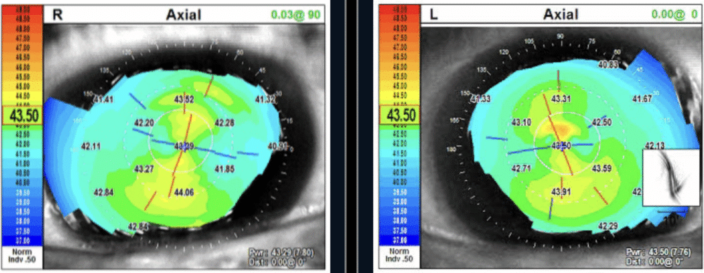
Corneal With-the-rule Astigmatism in both right and left eyes.
Corneal topography and aberrometry from OPD Scan III help your surgeon understand the exact curvature and regularity of the cornea, which is crucial when selecting the power and type of intraocular lens (IOL) for cataract surgery. Modern cataract care aims not only to remove the cloudy lens but also to reduce dependence on glasses.
For toric and premium IOLs, OPD Scan III identifies corneal astigmatism and maps higher-order aberrations, helping decide whether a toric lens is needed and candidacy for premium lifestyle IOLs such as Full Range of Vision or Extended Range of Vision IOLs.
Role in Keratoconus and corneal disorders3
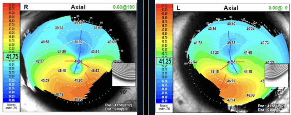
Pellucid Marginal Degeneration (a form of keratoconus) in both right eye left eyes.
OPD Scan III corneal topography maps corneal curvature, elevation, and irregularity, which are key for diagnosing and staging keratoconus and other ectatic corneal disorders. Research has demonstrated that its topographic and aberrometric indices show acceptable repeatability in mild and moderate keratoconus, making it useful for monitoring progression and post–corneal cross-linking follow-up.
Ocular Surface Disease and Dry Eye assessment4
Corneal topography from OPD Scan III can show subtle surface irregularities and tear-film–related distortion, helping identify ocular surface disease and dry eye that may not be obvious on basic exam alone. OPD is useful for assessing how ocular surface disease impacts vision supporting both diagnosis and monitoring symptoms such as fluctuation in vision from dry eye.
Dry eye can affect corneal curvature measurements which plays a critical role proper selection of intraocular lens for cataract surgery. Consequently, untreated dry eye is a common cause of dissatisfaction after cataract surgery, especially if refractive target is not met. A recent study found OPD Scan III among the most reliable devices for corneal curvature in dry eye patients, suggesting its measurements should be prioritized when planning cataract or refractive procedures.
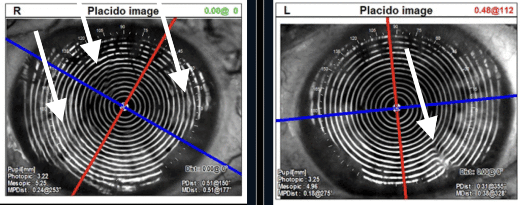
Arrows point to areas of surface irregularities of the tear film and/or corneal surface.
Why OPD Scan III matters for patients
For patients, OPD Scan III means more personalized care: your doctor can see not just how you see from the eye chart, but why you are seeing the way you do —by visualizing the full optical system and ocular surface. This information supports better cataract IOL selection, earlier detection of keratoconus and other corneal conditions, and more targeted dry eye management to protect both comfort and surgical outcomes.
Although this diagnostic test is not covered by OHIP, doctors at North Toronto Eye Care strongly recommend it as a crucial diagnostic aid for optimizing treatment plans and improving visual outcomes. By integrating multiple tests into one quick scan, OPD Scan III streamlines your visit while providing high-quality data aligned with modern practice patterns and evidence-based imaging strategies.
References
- https://files.advancingeyecare.com/Marco/opd_scan_iii.pdf
- https://www.guidelinecentral.com/guideline/10626/
- https://eyewiki.org/Corneal_Topography
- https://eyesoneyecare.com/resources/role-of-imaging-in-dry-eye-disease/